Mary Jones, Richard Fosbery, Dennis Taylor and, Jennifer Gregory Solutions for Chapter: Control and Coordination, Exercise 11: Question
Mary Jones Biology Solutions for Exercise - Mary Jones, Richard Fosbery, Dennis Taylor and, Jennifer Gregory Solutions for Chapter: Control and Coordination, Exercise 11: Question
Attempt the practice questions on Chapter 15: Control and Coordination, Exercise 11: Question with hints and solutions to strengthen your understanding. Biology for Cambridge International AS & A Level coursebook 2nd Edition Digital Access solutions are prepared by Experienced Embibe Experts.
Questions from Mary Jones, Richard Fosbery, Dennis Taylor and, Jennifer Gregory Solutions for Chapter: Control and Coordination, Exercise 11: Question with Hints & Solutions
LSs and TSs of striated muscle fibres were examined in an electron microscope. In the given picture shows drawings of the structures visible in a sarcomere in LS and TS as seen in a TEM. Explain why an electron microscope rather than a light microscope was used to study these sections.

Name the structures A, B, and C in the given figure.
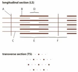
Name the regions of the sarcomere labelled D, E and F in the given figure.

State the region of the sarcomere where the TS was taken as shown in the given figure.

Explain how the sliding of filaments in a sarcomere leads to the contraction of a muscle fibre.