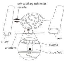
Suggest one role for the precapillary sphincter shown in the figure.
Important Questions on Transport in Mammals
The given image shows circulation in mammalian tissue.

With reference to this figure, describe the role of capillaries in the formation of tissue fluid.
Describe three ways in which tissue fluid differs from plasma.
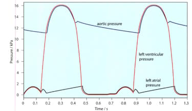
The given figure shows the pressure changes in the left atrium, left ventricle and aorta throughout two cardiac cycles.
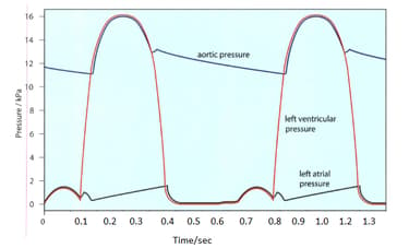
How long does one cardiac cycle last?
The given figure shows the pressure changes in the left atrium, left ventricle and aorta throughout two cardiac cycles.
What is the heart rate represented on this graph in beats per minute?
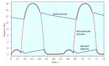
The given figure shows the pressure changes in the left atrium, left ventricle and aorta throughout two cardiac cycles. Make a copy of this diagram.

The contraction of the muscles in the ventricle wall causes the pressure inside the ventricle to rise. When the muscles relax, the pressure drops again.
In your copy of the diagram mark the time when the ventricle is contracting.
The given figure shows the pressure changes in the left atrium, left ventricle and aorta throughout two cardiac cycles. Make a copy of this diagram.
The contraction of the muscles in the ventricle wall causes the pressure inside the ventricle to rise. When the muscles relax, the pressure drops again.
In your copy of the diagram mark the time when the ventricle is relaxing.
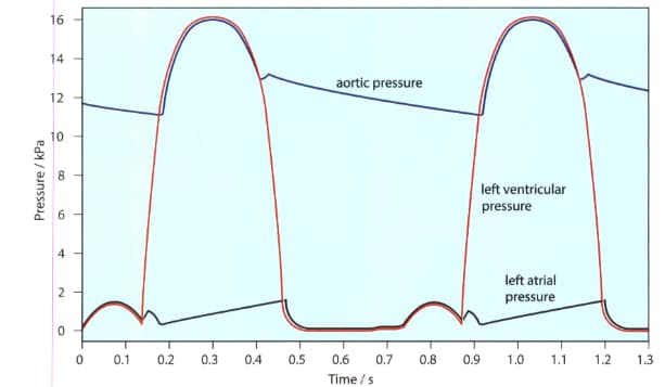
The given figure shows the pressure changes in the left atrium, left ventricle and aorta throughout two cardiac cycles. Make a copy of this diagram.

The contraction of muscles in the wall of the atrium raises the pressure inside it. This pressure also raised when the blood flows into the atrium from the veins, while the atrial walls are relaxed.
In your copy of the diagram mark the time when the atrium is contracting. (atrial systole)
The given figure shows the pressure changes in the left atrium, left ventricle and aorta throughout two cardiac cycles. Make a copy of this diagram.
The contraction of muscles in the wall of the atrium raises the pressure inside it. This pressure also raised when the blood flows into the atrium from the veins, while the atrial walls are relaxed.
In your copy of the diagram mark the time when the atrium is relaxing. (atrial diastole)