


Ellipse: Do you know the orbit of planets, moon, comets, and other heavenly bodies are elliptical? Mathematics defines an ellipse as a plane curve surrounding...
Last Modified 14-04-2025

Harvest Smarter Results!
Celebrate Baisakhi with smarter learning and steady progress.
Unlock discounts on all plans and grow your way to success!

Ellipse: Definition, Properties, Applications, Equation, Formulas
April 14, 2025
Altitude of a Triangle: Definition & Applications
April 14, 2025
Manufacturing of Sulphuric Acid by Contact Process
April 13, 2025
Refining or Purification of Impure Metals
April 13, 2025
Pollination and Outbreeding Devices: Definition, Types, Pollen Pistil Interaction
April 13, 2025
Acid Rain: Causes, Effects
April 10, 2025
Congruence of Triangles: Definition, Properties, Rules for Congruence
April 8, 2025
Complementary and Supplementary Angles: Definition, Examples
April 8, 2025
Nitro Compounds: Types, Synthesis, Properties and Uses
April 8, 2025
Bond Linking Monomers in Polymers: Biomolecules, Diagrams
April 8, 2025
Flagella and Cilia: Have you seen some microorganisms like Euglena and Paramoecium having hair-like or thread-like appendages that help the movement? The Flagella and Cilia are microscopic, contractile and filamentous processes of the cytoplasm capable of producing a current in the fluid medium for locomotion and passage of substances. Also, they act as sensory organs and perform many mechanical functions of the cell.
Cilia and flagella are structurally identical cell organelles that differ in length and function. Flagella can be found in bacteria and sperm cells, while cilia can be seen in species like Paramecium. Cilia are more numerous and shorter than flagella. This article will learn more about Flagella and Cilia differences with examples.

The cytoskeleton is a structure that helps the cells maintain their shape and internal organization. It also provides mechanical support that enables cells to carry out essential functions like division and movement.
These consist of the following types:
(i) Microtubules – These are unbranched, hollow tubules made up of tubulin protein. They contain \(13\) protofilaments and are \(25\,nm\) in diameter. They occur in centrioles, cilia/flagella, basal bodies, astral rays, spindle fibres, etc. They are non-contractile in nature.
(ii) Microfilaments – They are long, narrow, cylindrical rods made up of actin and myosin protein. They are contractile, solid structures with a diameter of about \(7\, nm.\)
Flagella (singular = flagellum) are long, hair-like structures that extend from the plasma membrane and are used in the movement of an entire cell.
Cilia (singular = cilium) are short, hair-like structures that are used to move entire cells or substances along the outer surface of the cell.
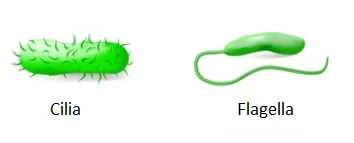
Fig: Cilia and Flagella
Eukaryotic and prokaryotic flagella are different in chemical composition and structure.
Both Flagella and Cilia are structurally similar and possess similar parts, i.e., basal body, rootlets, basal plate and shaft.
i. It is also known as Kinetosome or basal granule, or blepharoplasty. The basal body occurs embedded in the outer part of the cytoplasm below the plasma membrane.
ii. It is like a micro cylinder that has a structure similar to a centriole with nine triplet fibrils present on the periphery without a central fibril, even though a hub of protein is present here.
i. They are striated fibrillar outgrowths that develop from the outer lower part of the basal body.
ii. It helps in providing support to the basal body.
It is an area of high density which lies just above the basal body at the level of the plasma membrane.
i. The shaft is the hair-like projecting structure of the flagellum or cilium. The length is \(5\) to \(20\,\mu m\) in the case of the cilium and \(100\) to \(200\,\mu m\) in the case of the flagellum.
ii. Internally, it contains a semifluid matrix having an axoneme of \(9\) peripheral doublet fibrils and 2 central singlet fibrils. Hence, this arrangement is called \(9 + 2\) or \(11 – \)stranded.
iii. Each peripheral fibril consists of two microtubules. The sub-fibre A is slightly narrower, and it bears two bent arms, the outer one having a hook. They are about \(15\, nm\) long and made up of protein dynein with ATPase activity.
iv. The peripheral doublet fibrils are interconnected by \(A-B\) linkers of a protein called nexin between the \(B\)-sub fibre of one and the inner sidearm of the \(A\)-sub fibre of the adjacent fibril.
v. Each of their \(A\) sub-fibres sends a radial proteinaceous column to the centre. It is called radial spoke; the spokes are broader internally to form heads or knobs.
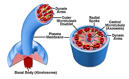
Fig: Structure of Flagella and Cilia
Cilia is of two types and are as follows:
Motile cilia are found on the cell surface in larger numbers. These are also found in the respiratory epithelium of the human respiratory tract and help by clearing the mucus or the dust particles out of the lungs.
Non-motile Cilia were first discovered in the year \(1898.\) These are long in structure and were believed to be a vestigial organelle. Recent studies and researchers presented the biological roles of primary cilia in that they function as a sensory cellular antenna that coordinates a large number of cellular signalling pathways.

The flagella structure and chemical composition are different in eukaryotes and prokaryotes. The bacterial flagella have the following features:
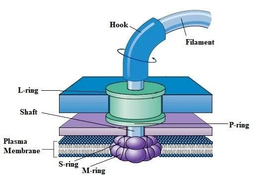
Fig: Structure of Prokaryotic Flagellum
Based on the number and arrangement of flagella, there are the following types of bacteria:
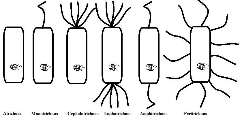
Fig: Arrangements of Flagella
| Eukaryotic Flagella | Bacterial Flagella |
| Enclosed is an outer membrane and consists of a pair of fibrils, surrounding a core of two fibrils. | Lacks a definite membrane. Composed of three longitudinal fibrils of flagellin protein twisted together into a helix that has a hollow core. |
| Whip back a fourth. | Rotatory motion. |
| Show \(9 + 2\) arrangement. | Do not have a \(9 + 2\) arrangement of protein filaments. |
| Tubulin and other types of protein filaments are present. | Flagellin proteins are mainly present. |
Some of the functions of flagella and cilia are as follows:
i. These help in locomotion in flagellated and ciliated organisms.
ii. The flagella or cilia also help capture food in many protozoans and some animals.
iii. They create water currents in certain aquatic animals for obtaining food.
iv. The flagella circulate food in the gastrovascular cavity of coelenterates.
v. The cilium of the respiratory tract helps to remove dust particles from it.
vi. The cilia in the alimentary canal of tunicates and lanceolate help in food movements and egestion.
vii. Motile cilia use their rhythmic undulation to sweep away substances, as in clearing dust, dirt, microorganisms and mucus, to prevent any diseases.
viii. Cilia plays an important role in the cell cycle and animal development, such as in the heart.
ix. Non-motile cilia serve as sensory apparatus for cells by detecting signals, and also they play crucial roles in sensory neurons.
x. Non-motile cilia can also be found in the kidneys to sense urine flow and the eyes of the photoreceptors of the retina.
xi. Flagella have an active role in aiding cell feeding and eukaryotic reproduction.
xii. In prokaryotes such as bacteria, flagella serve as propulsion mechanisms; they are the chief way bacteria swim through fluids.
xiii. Flagella also act as bridges or scaffolds for adhesion to host tissue.
The differences between cilia and flagella are:
| Cila | Flagella |
| They are numerous per cell (nearly \(300\) to \(4000\)). | They are few per cell (\(1\) to \(4\)). |
| They are smaller (nearly \(5\) to \(20\) micron). | They are comparatively larger, nearly \(100-200\) micron. |
| They usually occur throughout the surface of the cell. | They are commonly found at one end of the cell. |
| The surface is smooth. | The surface may contain flimmers. |
Through this article, we understood the structure and functions of flagella and cilia and the difference between them. These are the microscopic contractile and filamentous structure of the cytoplasm. It creates food currents, acts as sensory organs and performs many mechanical functions of the cell. Cilia and Flagella are identical structures, but both can be distinguished by their number and function.
Important Questions on Flagella and Cilia

Q.1. Write one function of cilia.
Ans: The cilia also help in capturing food in many protozoans and some animals. They also help in feeding, locomotion, aeration, circulation, etc.
Q.2. Do all bacteria have flagella?
Ans: Flagella occur on both Gram-positive and Gram-negative bacteria, and their presence can be useful in identification. For example, they are found on many species of bacilli but rarely on cocci. E. coli has flagella all over the body.
Q.3. What are flagella made of?
Ans: Flagella are composed of subunits of a low-molecular-weight protein called flagellin that is arranged in a helical manner.
Q.4. What are the types of flagella?
Ans: There are two main types of flagella in eukaryotes:
1. Whiplash flagellum is one that does not have hairy flimmers on the surface.
2. The tinsel flagellum is one that has lateral hair-like projections or flimmers, or mastigonemes on the surface.
Q.5. Where are flagella found in the human body?
Ans: In humans, the flagellum is only seen in gametes, i.e., in sperms that help to swim towards the ovum.
We hope this detailed article on Flagella and Cilia helps you in your preparation. If you get stuck do let us know in the comments section below and we will get back to you at the earliest.


